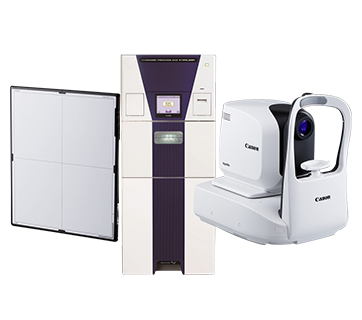
Xephilio OCT-R1
Xephilio OCT-R1

Xephilio OCT-R1
Introducing a powerful combination of a fully automated SD-OCT with unsurpassed HD resolution retinal imaging technology made by Canon.
Promotion
Key Features
Features
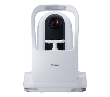
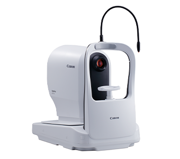
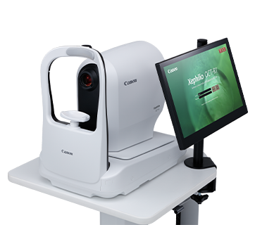
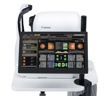
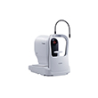
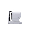
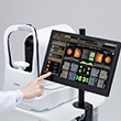
Full Auto OCT + Retinal Camera
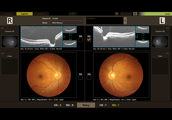
The combination of OCT and high-definition retinal camera in the Xephilio OCT-R1 makes it an ideal solution for screening and practices in high patient volume. The OCT-R1 offers a large and high-quality OCT scan up to 14.7 x 13.4 mm. Combined with the retinal camera (24.2MP resolution), the OCT-R1 produces high definition and unsurpassed true-color imaging. The comprehensive RX software is designed to automatically combine the information from both the OCT and the retinal camera for complete reporting and diagnostic support.
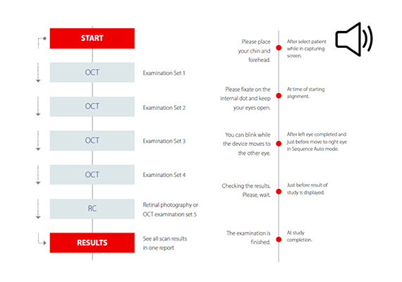
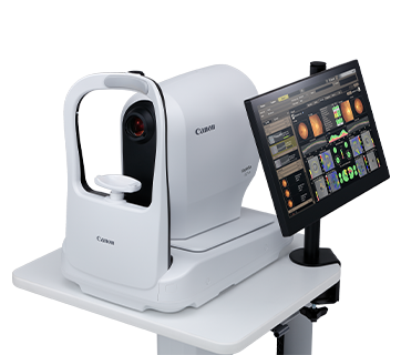
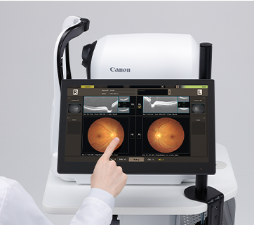
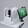
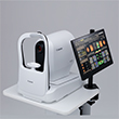
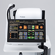
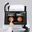
Sequential Auto and Audio Guidance
The OCT-R1 offers a fully automated examination protocol that can be initiated with just one touch. This advanced feature streamlines the imaging process and ensures accuracy and consistency in the imaging data obtained from both eyes. When used in conjunction with the audio guidance feature, this makes the device exceptionally easy to use for both patients and operators. The OCT-R1 is an excellent choice for practitioners who value speed, accuracy, and patient comfort.
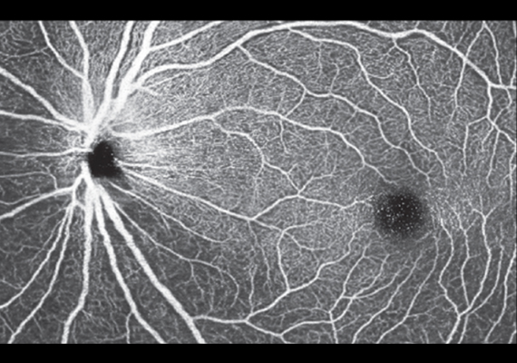
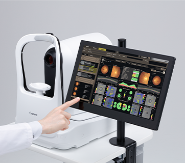
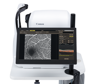
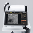
OCT Angiography and Angio Expert
The Xephilio OCT-R1 allows clinicians to visualise the microvasculature of the retina in extraordinary detail. The movement of the red blood cells in the retinal vasculature can be detected with the optional Angio Expert software, making it possible to identify the tiniest vessels with precision in both 2D and 3D format within a few seconds. Various scan areas, ranging from 3 x 3 up to 13.4 x 13.4 mm (A-scan x B-scan), are possible for comprehensive analysis. Furthermore, injection with fluorescein or dilation of the pupil is not required, making it patient-friendly and hassle-free.
Specifications
Specifications
|
OCT Type |
Spectral Domain |
|
Light Source |
SLED |
|
Central OCT Wavelength |
880 nm |
|
Axial Resolution (Optical) |
7μm |
|
Scan Width |
3 ~ 14.7 mm |
|
Scan Depth |
2.3 mm |
|
Scan Rate |
50,000 A scans/sec |
|
Minimum Pupil: OCT |
2.5 mm |
|
Retinal Observation |
Digital Camera (IR image) |
|
Photography |
Colour, Red free, Cobalt, Anterior segment |
|
Photography Angle |
45 degrees / 30 degrees digital |
|
Resolution |
24.2 MP / Centre resolution: 63 lines |
|
Minimum Pupil Camera |
4.0 mm (3.3 SP) |
|
Chinrest |
Motorised |
|
Dimension and Weight |
|
|
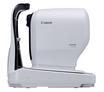
Dimension (W x D x H) |
335 x 490 x 473 mm |
|
Weight |
23 kg |
|
OCT Scan Parameters |
|
|
A-scan x B-scan |
|
|
Macula 3D |
1,024 x 128 |
|
Glaucoma 3D |
1,024 x 128 |
|
Disc 3D |
512 x 256 |
|
Wide 3D |
512 x 128 |
|
Custom 3D |
1,024 x 128 |
|
Cross |
1,024 x 1 x 2 |
|
Multicross |
1,024 x 1 x 2 |
|
Radial |
1,024 x 12 |
|
Scan Width (V x H) (mm) |
|
|
Macula 3D |
10 x 10 |
|
Glaucoma 3D |
10 x 10 |
|
Disc 3D |
6 x 6 |
|
Wide 3D |
13 x 10 |
|
Custom 3D |
3 x 3 ~ 14.7 x 13.4 |
|
Cross |
3 x 3 ~ 14.7 x 13.4 |
|
Multicross |
3 x 3 ~ 14.7 x 13.4 |
|
Radial |
3 x 3 ~ 14.7 x 13.4 |
|
Scan Direction |
|
|
Macula 3D |
Horizontal |
|
Glaucoma 3D |
Vertical |
|
Disc 3D |
Horizontal |
|
Wide 3D |
Vertical / Horizontal |
|
Custom 3D |
Horizontal or Vertical |
|
Cross |
Vertical / Horizontal |
|
Multicross |
Vertical / Horizontal |
|
Radial |
Radial |
|
Averaging |
|
|
Cross |
1/5/10/20/50 |
|
Multicross |
1/5/10/20 |
|
Radial |
1/5/10/20 |
*Optional:
- OCT Angiography (Angio Expert Software)
- Intelligent De-noise
- Retinal Expert (RX) Viewer
- Retinal Expert (RX) Server
- Mosaic / Panoramic Software
- EL-1 Fixation Target
Photo Library
Product Disclaimer
- 01. Please refer to individual country / region websites and respective sales offices for product availability.
- 02. Specifications, availability and terms of offers may change without notice.
- 03. Products / Services may be manufactured by and/or supplied to us by third party manufacturers / suppliers for distribution / resale (non-Canon brand products).